Bursitis
Every joint in the body has bursa around them, with the body containing over 150 bursar.
A bursa is a small fluid filled sac that acts as a cushion between two surfaces to reduce friction- e.g. between bone and an overlying tendon, between tendon and overlying skin, or between bone and skin.
If the bursa comes inflamed this is termed a bursitis.
In the knee the three common bursa that cause symptoms are the pre patellar bursa, the infra-patellar bursa and the pes anserinus bursa
Pre patellar bursitis
Inflammation of the pre patellar bursa (pre patellar bursitis) is also commonly known as 'housemaids knee'.
The pre patellar bursa lies over the knee cap.
Jobs which involve frequent kneeling and the risk of cuts over the kneecap are associated with inflammation of the pre patellar bursa.
In some cases the bursa can become infected.
Infra patellar bursitis
The infra patellar bursa can be split into two parts: superficial and deep.
The superficial bursa lies between the skin and the tibial tuberosity.
The deep bursa lies behind the patella tendon and in front of the proximal tibia.
Pes anserinus bursitis
The pes anserinus bursa separates the sartorial, gracilis and semi-tendinosus tendons from the underlying medial collateral ligament and bony surface of the tibia.
Signs and symptoms
The site of the the bursitis is normally red, inflamed, swollen and tender to palpation.
Diagnosis of bursitis
The diagnosis of bursitis will be based on a clinical history and examination.
An occupation, sport or hobby associated with frequent kneeling or a recent trauma to the site of a bursa may be risk factors.
As mentioned the site of the bursa is often red, inflamed, swollen and tender to palpation.
A radiograph may be performed in the event of a trauma or a history of osteoarthritis.
Ultrasound is very useful in the diagnosis of bursitis.
In some circumstances an MRI scan may be performed.
Differential diagnosis
It is important to exclude an infected bursa from an inflamed bursa.
It is also important to exclude an infection of the associated knee joint.
Treatment
The treatment of an acute bursitis normally requires rest of the knee joint, with a short course of paracetamol, anti-inflammatory medications and intermittent ice packs. A brace may be used to help rest the knee joint.
In long standing cases an ultrasound guided steroid injection can be performed to reduce inflammtion.
Modification of activities causing the bursitis can help future prevention.
Jobs requiring frequent kneeling may benefit from the use of knee pads.
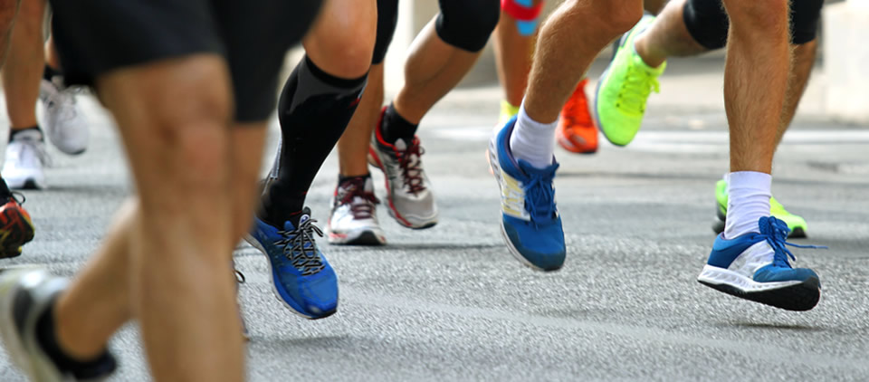
Sporting activities may require sport specific rehabilitation (with a focus on technique, form and training) and a biomechanical assessment (iliotibial bursitis can occur with running and may require assessment of foot shape and appropriate footwear).
In rare circumstances a chronic, recurrent bursitis may require surgical excision of the bursa.
An acute infected bursitis may require oral or intravenous antibiotics and in some cases a surgical drainage of the infection.